doi: 10.56294/hl2024.188
ORIGINAL
Analysis and prediction of different stages of brain tumor from mri using ai models
Análisis y predicción de diferentes estadios de tumores cerebrales a partir de resonancias magnéticas mediante modelos de inteligencia artificial
Rustam
Navruzov1 ![]() *,
Jasur Ismoilov2
*,
Jasur Ismoilov2 ![]() *,
Mukhammadshokir Bahadirkhanov3
*,
Mukhammadshokir Bahadirkhanov3
![]() *, Umida Almatova4
*, Umida Almatova4
![]() *,
Jamolbek Djuraev5
*,
Jamolbek Djuraev5 ![]() *, Alisher Mikhiddinov6
*, Alisher Mikhiddinov6
![]() *,
Nadira Mirametova7
*,
Nadira Mirametova7 ![]() *
*
1Bukhara State Medical Institute named after Abu Ali ibn Sino. Bukhara, Uzbekistan.
2Samarkand State Medical University. Samarkand, Uzbekistan.
3Republican Research Centre of Emergency Medicine. Uzbekistan.
4Jizzakh State Pedagogical University. Uzbekistan.
5Tashkent Medical Academy. Uzbekistan
6Termez Branch of Tashkent Medical Academy. Uzbekistan.
7Ajiniyaz Nukus State Pedagogical Institute.
Cite as: Navruzov R, Ismoilov J, Bahadirkhanov M, Almatova U, Djuraev J, Mikhiddinov A, et al. Analysis and prediction of different stages of brain tumor from mri using ai models. Health Leadership and Quality of Life. 2024; 3:.188. https://doi.org/10.56294/hl2024.188
Submitted: 29-02-2024 Revised: 19-06-2024 Accepted: 15-10-2024 Published: 16-10-2024
Editor: PhD.
Prof. Neela Satheesh ![]()
Corresponding Author: Rustam Navruzov *
ABSTRACT
Medical imaging uses computer techniques to create images of the human body’s inside for diagnostic purposes. Image segmentation is crucial for many medical assessments, navigation, surgical planning, and diagnosis. There are now three techniques available for segmenting the region of interest: manual, semi-automated, and automatic. In this paper, a hybrid way to deal with the identification and characterization of brain cancers utilizing attractive reverberation imaging is proposed. It is very challenging to analyze brain cancers due to the varieties in growth position, structure, and size. The fundamental target of this study is to provide specialists with an exhaustive assortment of writing on the use of attractive reverberation imaging (MR) to the identification of cerebrum diseases. This study proposed a number of techniques for the statistical image processing and artificial intelligence-based tumor and brain cancer diagnosis. This concentrate likewise incorporates an evaluation grid for a particular framework utilizing explicit frameworks and dataset types. At last, our review gathers every one of the information expected to analyze and figure out malignant growths, including their advantages, disadvantages, headways, and possible future patterns.
Keywords: Brain Tumor; Classification; Segmentation; Deep Learning; Machine Learning.
RESUMEN
La imagen médica utiliza técnicas informáticas para crear imágenes del interior del cuerpo humano con fines diagnósticos. La segmentación de imágenes es crucial para muchas evaluaciones médicas, la navegación, la planificación quirúrgica y el diagnóstico. Actualmente existen tres técnicas para segmentar la región de interés: manual, semiautomática y automática. En este trabajo se propone una forma híbrida de abordar la identificación y caracterización de los cánceres cerebrales utilizando atractivas imágenes reverberantes. Es muy difícil analizar los cánceres cerebrales debido a las diferencias en la posición de crecimiento, la estructura y el tamaño. El objetivo fundamental de este estudio es proporcionar a los especialistas un surtido exhaustivo de escritos sobre el uso de imágenes reverberantes atractivas (MR) para la identificación de enfermedades cerebrales. Este estudio propone una serie de técnicas para el procesamiento estadístico de imágenes y el diagnóstico de tumores y cáncer cerebral basado en inteligencia artificial. Este concentrado incorpora asimismo una tabla de evaluación para un marco particular utilizando marcos explícitos y tipos de conjuntos de datos. Por último, nuestra revisión reúne toda la información esperada para analizar y averiguar crecimientos malignos, incluyendo sus ventajas, desventajas, avances y posibles patrones futuros.
Palabras clave: Tumor Cerebral; Clasificación; Segmentación; Deep Learning; Machine Learning.
INTRODUCTION
Brain tumours and their investigation are becoming increasingly intriguing due to the expanding field of IP technologies. As indicated by the Public Brain Cancer Establishment’s evaluation, the frequency of cerebrum growths among masses and the demise rate from cerebrum growths are on the ascent universally.(1,2) Every five years, data on the survival of patients with brain tumours are gathered (last collected in January 2020). The field of brain tumours has also been highlighted by a number of researches over the past few decades, which may or may not be followed by steps like outcome prediction, therapy planning, and classification.(3) Noise, absent boundaries, and insufficient contrast are common factors that affect medical picture segmentation.(4) These problems are successfully managed by diagnostic imaging techniques like magnetic resonance imaging (MRI) and computed tomography (CT) scans. Numerous ailments can be diagnosed with the help of these imaging techniques.(5,6) Because magnetic resonance imaging (MRI) makes use of safe magnetic fields and radio waves, it is a popular tool for the effective diagnosis and treatment of brain tumours.(7)
Brain Tumor - Overview
MR images can be processed fully automatically, semi-automatically, or manually with human input. Medical image processing requires accurate segmentation, which is often completed by hand by specialists and requires a significant amount of time.(8) It will take some time to develop a segmentation approach that is both effective and fully automatic. Furthermore, a second viewpoint is always necessary because, in these systems, human life is the main concern.(9) Indeed, even without any specialists, the viability of mechanized procedures relies just upon the information bases. Specialists have proposed various strategies to further develop these information bases and, thusly, the viability of cancer discovery systems.(10) The level of client oversight and calculation ease are the main boundaries that influence the clinical and neurotic acknowledgment of division draws near.(11) The requirement for complete client oversight that accompanies manual strategies makes a push for improved arrangements. The self-loader or intelligent methodologies that are right now being used will win practically speaking from here on out.(12) In clinical contexts, radiologists commonly employ semi-automated segmentation approaches to substitute the time-consuming and challenging human segmentation processes.(13) User initialisation occurs in almost all semi-automatic processes, and accuracy requires frequent user interaction. In light of this, the work included in this thesis also tries to envision how these automatic/semiautomatic systems will be accepted and expand in the medical and scientific domains for a variety of tumor classes in figure 1.
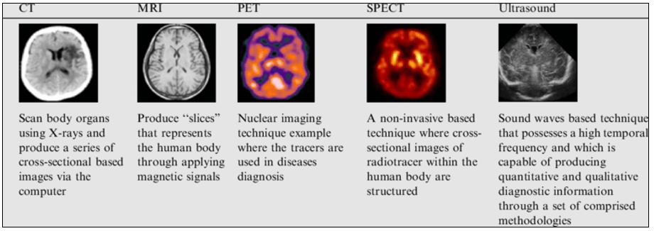
Figure 1. Imaging modalities used for the diagnosis of brain tumor
Tumor Types and their Grades
With a focus on MR imaging, the goal of this study is to thoroughly investigate multi-grade brain tumour detection techniques. It has been observed that the performance of the segmentation or classification strategy tends to follow a certain pattern based on the type of tumour present in the collected dataset or produced brain images.(14,15) The World Health Organization's (WHO) stringent histopathological grading guidelines for classifying brain tumours complicate medical imaging diagnostic and detection techniques. In addition to these tumour kinds, lesions and Alzheimer's disease are also considered based on their data sources and detection techniques. While bearing this constraint in brain, numerous academics are making progress in the development of computer assisted detection (CAD).(16)
METHOD
Pre – processing technique
Pre-processing is the process of converting an original image to grayscale in order to minimize noise and remove undesired elements for image enhancement and reconstruction. The grayscale image's intensity is either 1 or 0 at every location.(17) Each pixel has an intensity value that is devoid of color. Fractions may also be used to express intensity values. By providing exact information, it aids in the segmentation process. Either a high pass or low pass filter type is available. Several kinds of filters have been employed by the various researchers to remove noise from the image. In the research project, Deepthi Murthy used the Sobel filter. The Sobel filter is a sort of derivative mask that operates on the basis of calculating pixel intensity differences.
It is possible to eliminate Poisson noise and picture de-noising from the. It makes use of the window that slides across the picture. During the process, the pixel output intensity value in the window is determined by taking the pixel median intensity value. It lowers noise while maintaining the image's edges.
Detection and identification of lesions
Because of factors including lesion irregularity, differences in measurement between and within observers, and different scanning levels derived from different analyses, the actual estimate of tumor size cannot be based just on lesion diameter. Brain tumors can vary widely in size, shape, and location.(18) Experts trace the lesion manually as part of the standard procedure. The lesion's automatic identification reduces the amount of time that practitioners need to spend on analysis.
Feature selection
An artificial bee colony (ABC) based feature selection (FS) algorithm i.e. ABC-FS to determine the appropriate features for stroke prediction. In literature, feature selection algorithm can either implement in supervised or unsupervised manner.(19,20) In this work, unsupervised approach is used for implementing the ABC-FS algorithm. This approach determines the optimal clusters initially, then weight of each feature is computed on the basis of optimal centroid. The relevancy of features is described using the weight function. In turn, higher weight function corresponds to more relevant feature. Other side, artificial bee colony is a meta-heuristic algorithm based on the bee behaviour especially characteristic of honey bees for determining the nectar amount. The employed and onlooker bees responsible for exploration as well as exploitation of solution in search space, while scout be responsible for exploration search space for good candidate solution.
Brain Tumor Classification Module
Since there are various tasks involved in the classification, it is ineffective to use a different model for each activity. Different approaches are taken to classify the tumor from the same brain MRI imaging.(21) It is an intelligent paradigm of machine learning that can widely adopted for handling variety of problems and defined using complex relationships and multiple representation levels. Further, both of feature extraction and classification tasks are handled effectively using deep learning technique.
CNN Model
The most advanced technique for identifying brain tumors in medical pictures is to use CNNs. The CNN model does not consider the corresponding masks and instead only uses MRI scans for tumor prediction. This suggests that the CNN does not use the associated masks' additional information, when predicting the presence or characteristics of tumors based solely on its analysis of the MRI images.(22,23) CNN model considers the feature extracted through ABC-FS algorithm as input to model. The main layer of CNN model is convolutional layer and this layer process the input data using different kernels. The kernels are represented through features of dataset. The aim of this work is to generate the feature map that can be acted as input for next layer This layer considers the feature map as input and aim of this layer is to validate the accurate information that can transfer to the next layer. In other words, this layer validates the output of the convolutional layer in terms of features through consecutive operations. Further, this layer is likewise tended to the overfitting issue of information. An actuation capability is additionally viewed as on this layer for bit improved results and the errand of enactment capability is to initiate the neurons. In addition, the actuation of neurons is relied upon the premise of data, assuming data is proper, comparing neuron can enact in any case it remain deactivate.(24,25) A neuron value is also computed for each neuron through activation function. This work considers the Tanh function as activation function for max-pooling layer in figure 2. This layer considers all extracted feature and combined these features for generating a final feature set.
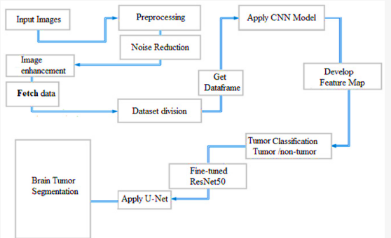
Figure 2. Proposed model architecture
ResNet50 Model
The standard strategy for BT location and grouping utilizing MRI involves using CNN to refine the ResNet50 model. Utilizing the huge ImageNet dataset, a CNN model known as ResNet50 was prepared for object recognizable proof errands. It is comprised of various layers, including convolutional, pooling, and completely connected layers.(26) This model's utilization as an element extractor can assist with the errand of distinguishing brain growths. The most minimal layers of the ResNet50 model concentrate highlights from freely accessible photographs that could be valuable for cerebrum cancer recognizable proof. The last couple of levels of the ResNet50 model are supplanted with another arrangement of completely connected layers explicitly intended for the errand of brain growth ID and order.
At the point when the extra completely associated layers are added, the model all in all can be upgraded utilizing a new dataset of MRI pictures.(27,28) To refresh the loads of each layer in the model, stochastic angle plunge and backpropagation are utilized. The info information comprises of brain MRI filters, which are frequently preprocessed to upgrade contrast between the cancer and encompassing tissue. The reexamined model yields a likelihood conveyance over the class names growth (yes) or non-cancer (no). This probability can then have a limit set to it to survey on the off chance that the growth is available or not.
By extricating discriminative properties during preparing, the superior ResNet50 model with CNN figures out how to recognize growths from sound brain MRI information. The pre-prepared ResNet50 model offers a vigorous introductory arrangement of highlights for cerebrum cancer location, and it very well might be changed on a new dataset of MRI pictures to make the model more intended for the main job. This approach has shown extremely exact outcomes for BT location and categorisation utilizing MRIs.
RESULTS
We experimented with the tumor identification and classification modules by changing hyperparameters such learning rate, batch, and epochs. The hyperparameters that yield superior outcomes were selected. Twenty percent of the data are used to test the trained modules. The multi-task classifier's task-specific individual layers each have distinct loss computations that are later integrated. A representative outcome is displayed in figure 3. This model's total accuracy was 92 %. The tumour identification module is evaluated using the Dice coefficient. An average Dice score of 0,89 is obtained.
When matched with further developed ResNet50 and U-Net, convolutional brain network demonstrating can be a strong strategy for thorough brain growth recognizable proof, grouping, and division. This technique boosts the qualities of each model to work on all out precision.
CNN Model Results
The CNN configuration has shown promising results in this work with regards to brain cancer finding. The model showed great degrees of measurable exactness, accuracy, review, and F1 score, making it a solid instrument for diagnosing brain cancers from MRIs. This model was prepared utilizing a cerebrum MRI dataset that contained named growth and non-cancer pictures. The CNN model accomplished a high exactness of 92 % in cerebrum growth finding, as shown by the disarray lattice in figure 3. Figure 4 red and blue lines likewise offer a visual portrayal of exactness and misfortune. The extent of right certain recognitions to all sure identifications is shown by the accuracy number, which shifts from 90 % to 94 %. This figure was likewise high.
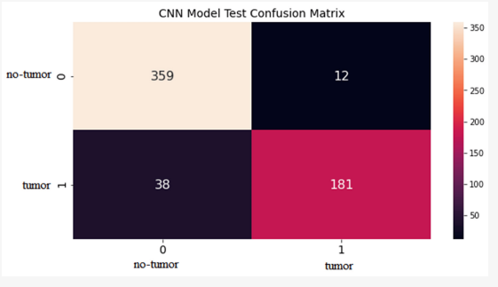
Figure 3. Confusion matrix results
Due to its solid measurable qualities, the CNN model can be utilized by radiologists and specialists to rapidly and successfully analyze cerebrum growths from MRI filters. Clinical faculty can treat patients all the more quickly and really when brain growths are recognized early.
The proposed model purposes the CNN model with further developed ResNet50 engineering to order brain growths. The model is prepared utilizing a dataset of named growth sorts from cerebrum MRI checks, which incorporates both cancer and non-cancer pictures. The red and blue lines in the disarray grid in fig. show the high precision, generally 94 %, of the ResNet50 model's brain cancer grouping results. The model's precision, which finds out the extent of precisely grouped positive growth types among every single positive characterization, is similarly high, commonly going from 93 % to 96 %. Review is major areas of strength for another; it ordinarily goes from 87 % to 98 %. Review evaluates the extent of genuine positive growth type characterizations out of all real cancer type cases.
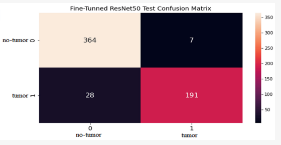
Figure 4. Final results of Confusion matrix
CONCLUSIONS
This work proposed a CNN model that consolidated better variants of ResNet50 and U-net for the distinguishing proof and categorisation of brain growths in MRI information. This model joins the best parts of two distinct designs to achieve high exactness in the two assignments. The Calibrated ResNet50 engineering is utilized to distinguish brain growths or identify the presence of a cancer in MRIs. The U-net engineering is utilized to accomplish brain growth division, which is the course of exactly secluding the cancer from the encompassing solid tissue. The review evaluated CNN and improved ResNet50 as two models' characterization and growth identification capacities utilizing MRI pictures. That's what the discoveries show, concerning factual qualities for both cancer and non-growth groupings, the refreshed ResNet50 model beats the CNN model. Following changes, the superior ResNet50 model delivered results for the non-cancer class of 0,98 accuracy, 0,95 review, 0,93 F1 score, and 0,94 exactness, and for the growth class of 0,87 accuracy, 0,92 review, 0,88 F1 score, and 0,96 precision. The accompanying results were acquired from the adjusted ResNet50 model: SI: 0,95, DSC: 0,91, and IoU: 0,91. These discoveries propose that the improved ResNet50 model could be a useful device for precisely distinguishing and ordering brain tumor growths from MRI.
BIBLIOGRAPHIC REFERENCES
1. Machiraju Jaya Lakshmi & S.Nagaraja Rao., “Brain tumor magnetic resonance image classification: a deep learning approach,” Soft Computing, vol. 26, pp.6245–6253, 2022, doi: https://doi:org/10.1007/s00500-022-07163-z.
2. Wen Jun & Zheng Liyuan, “Brain Tumor Classification Based on Attention Guided Deep Learning Model,” International Journal of Computational Intelligence Systems, vol.15, pp. 35, 2022, doi: https://doi:org/10.1007/s44196-022-00090-9.
3. Arshia Rehman et.al., “A Deep Learning-Based Framework for Automatic Brain Tumors Classification Using Transfer Learning.” Circuits Systems and Signal Processing, vol. 39, pp. 757–775, 2020, doi: https://doi:org/10.1007/s00034-019-01246-3.
4. Fernando, T. et.al., “Deep Learning for Medical Anomaly Detection – A Survey,” ACM Computing Surveys, vol. 54, issue (7), pp. 1-37, 2021, doi: https://doi:org/10.1145/3464423.
5. Alexander Selvikvåg Lundervold & Arvid Lundervold, “An overview of deep learning in medical imaging focusing on MRI,” Zeitschrift für Medizinische Physik, vol. 29, issue (2), 2019, pp. 102-127, doi: https://doi:10.1016/j.zemedi.2018.11.002.
6. Rundo, L. et al., “Semi-automatic Brain Lesion Segmentation in Gamma Knife Treatments Using an Unsupervised Fuzzy C-Means Clustering Technique,” Smart Innovation, Systems and Technologies, vol 54. Springer, Cham. 2016, doi: https://doi:org/10.1007/978-3-319-33747-0_2.
7. Stijn Bonte et al., “Machine learning based brain tumour segmentation on limited data using local texture and abnormality,” Comput Biol Med, vol. 98, pp. 39-47, 2018, doi: https://doi:10.1016/j.compbiomed.2018.05.005
8. Mohsen, Heba, et al. Classification using deep learning neural networks for brain tumors. Future Computing and Informatics Journal, Vol 3, pp 68-71, 2018. DOI: 10.1016/j.fcij.2017.12.001.
9. Sobhaninia, Zahra, et al. Brain Tumor Segmentation Using Deep Learning by Type Specific Sorting of Images. arXiv preprint arXiv:1809.07786 (2018).
10. Widhiarso, Wijang, Yohannes Yohannes, and Cendy Prakarsah. Brain Tumor Classification Using Gray Level Co-occurrence Matrix and Convolutional Neural Network. IJEIS (Indonesian Journal of Electronics and Instrumentation Systems), Vol 8, pp 179-190, 2018. DOI: 10.22146/ijeis.34713.
11. Chandra, Saroj Kumar, and Manish Kumar Bajpai. Effective algorithm for benign brain tumor detection using fractional calculus. TENCON 2018-2018 IEEE Region 10 Conference. IEEE, 2018. DOI: 10.1109/TENCON.2018.8650163.
12. Seetha, J., and S. S. Raja. Brain Tumor Classification Using Convolutional Neural Networks. Biomedical & Pharmacology Journal, Vol 11, pp 1457-1461, 2018. DOI: 10.1007/978-981-10-9035-6_33.
13. Cheng, Jun, et al. Enhanced performance of brain tumor classification via tumor region augmentation and partition. PloS one, Vol 10, 2015. DOI: 10.1371/ journal.pone.0140381.
14. Sajna, V., & Dharmaraj, A. (2024). Awareness of Central Sector Scheme among the Entrepreneurs in MSME Sector: An Investigative Study. Indian Journal of Information Sources and Services, 14(3), 115–122. https://doi.org/10.51983/ijiss-2024.14.3.16
15. Babenko, V., Danilov, A., Vasenin, D., & Krysanov, V. (2021). Parametric Optimization of the Structure of Controlled High-voltage Capacitor Batteries. Archives for Technical Sciences, 1(24), 9–16. https://doi.org/10.7251/afts.2021.1324.009B
16. Turan, F., & Ergenler, A. (2021). DNA Damage in Hybrid Tilapia (Oreochromis niloticus x O. aureus) Exposed to Short-Transport Process. Natural and Engineering Sciences, 6(3), 190-196. http://doi.org/10.28978/nesciences.1036849
17. Faisal, T., & Dharmaraj, A. (2024). Perception of E-learning among the Students Studying in Higher Education Institutions with Reference to Kerala. Indian Journal of Information Sources and Services, 14(4), 66–72. https://doi.org/10.51983/ijiss-2024.14.4.11
18. Cvijić, R., & Milošević, A. (2020). Reproduction of the Mineral – Raw Material Base problems in the Republic Srpska. Archives for Technical Sciences, 2(23), 1–8.
19. Min, S., & Atan, N. A. (2024). Students’ Performance and Perceptions of Authentic E-Learning Activities in English Intercultural Learning. Indian Journal of Information Sources and Services, 14(4), 92–102. https://doi.org/10.51983/ijiss-2024.14.4.15
20. Aydın, M., & Karadurmuş, U. (2021). The new maximum length of the Solea solea (Linnaeus, 1758) in the Turkish Coast of Black Sea. Natural and Engineering Sciences, 6(3), 261-264. http://doi.org/10.28978/nesciences.1036856
21. Danková, Z., Štyriaková, I., Kovaničová, Ľ., Čechovská, K., Košuth, M., Šuba, J., Nováková, J., Konečný, P., Tuček, Ľ., Žecová, K., Lenhardtová, E., & Németh, Z. (2021). Chemical Leaching of Contaminated Soil – Case Study. Archives for Technical Sciences, 1(24), 65–72.
22. Agina-Obu, R., & Oyinkepreye Evelyn, S.-G. (2023). Evaluation of Users’ Satisfaction of Information Resources in University Libraries in Nigeria: A Case Study. Indian Journal of Information Sources and Services, 13(1), 1–5. https://doi.org/10.51983/ijiss-2023.13.1.3378
23. Lukić, M. (2019). An Analysis of the Influence of Air Temperature and Humidity on Outdoor Thermal Comfort in Belgrade (Serbia) Using A Simple Heat Index. Archives for Technical Sciences, 2(21), 75–84.
24. Eiriemiokhale, K., & James, J. B. (2023). Application of the Internet of Things for Quality Service Delivery in Nigerian University Libraries. Indian Journal of Information Sources and Services, 13(1), 17–25. https://doi.org/10.51983/ijiss-2023.13.1.3463
25. Đurić, N., & Đurić, D. (2019). Landslides on the Roads of the Northern Part of the Republic of Srpska as a Result of Elemental or Anthropogenic Processes. Archives for Technical Sciences, 2(21), 11–24.
26. Salokhiddinov, A., et al. (2020). Impact of climate change on irrigated agriculture in steppe zones of Uzbekistan. IOP Conference Series: Materials Science and Engineering, 883, 012073. https://doi.org/10.1088/1757-899X/883/1/012073
27. Karimov, B. K., et al. (2020). Relationship between the concentrations of nitrogen compounds and the water discharge in the Chirchiq River, Uzbekistan. IOP Conference Series: Earth and Environmental Science, 614, 012154. https://doi.org/10.1088/1755-1315/614/1/012154
28. Karimov, N., et al. (2024). Exploring food processing in natural science education: Practical applications and pedagogical techniques. Natural and Engineering Sciences, 9(2), 359-375. https://doi.org/10.28978/nesciences.1574453
FINANCING
No financing.
CONFLICT OF INTEREST
The authors declare that there is no conflict of interest.
AUTHORSHIP CONTRIBUTION
Data curation: Rustam Navruzov, Jasur Ismoilov, Mukhammadshokir Bahadirkhanov, Umida Almatova, Jamolbek Djuraev, Alisher Mikhiddinov, Nadira Mirametova.
Methodology: Rustam Navruzov, Jasur Ismoilov, Mukhammadshokir Bahadirkhanov, Umida Almatova, Jamolbek Djuraev, Alisher Mikhiddinov, Nadira Mirametova.
Software: Rustam Navruzov, Jasur Ismoilov, Mukhammadshokir Bahadirkhanov, Umida Almatova, Jamolbek Djuraev, Alisher Mikhiddinov, Nadira Mirametova.
Drafting - original draft: Rustam Navruzov, Jasur Ismoilov, Mukhammadshokir Bahadirkhanov, Umida Almatova, Jamolbek Djuraev, Alisher Mikhiddinov, Nadira Mirametova.
Writing - proofreading and editing: Rustam Navruzov, Jasur Ismoilov, Mukhammadshokir Bahadirkhanov, Umida Almatova, Jamolbek Djuraev, Alisher Mikhiddinov, Nadira Mirametova.